 Get Insights

Accurate Ultrasound Image Assessment: Key Metrics, Artifacts, and Review Criteria
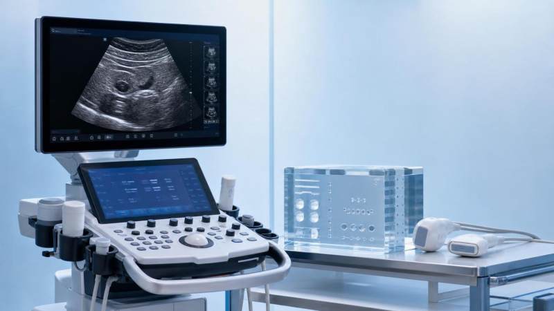
Accurate ultrasound image assessment matters because modern systems can produce images that look convincing while still missing technical consistency. In procurement, validation, and comparative review, visual plausibility is not enough. What matters is whether image quality holds across depth, tissue types, presets, probes, and operating conditions, and whether performance claims stand up to repeatable measurement.
That question has become more urgent as healthcare shifts toward value-based purchasing and digitally connected workflows. Ultrasound platforms now combine beamforming, post-processing, AI-assisted features, and software-defined controls. The result is a wider gap between a polished demo and dependable clinical-grade output. Accurate ultrasound image assessment closes that gap by linking image appearance to engineering integrity, usability, compliance expectations, and long-term service value.
Why image assessment now requires more than a visual check
An ultrasound image is the end product of many interacting variables. Transducer design, channel count, focusing strategy, signal processing, thermal management, and display mapping all influence the final result.
A system may look sharp in one preset and degrade in another. It may perform well at shallow depths yet lose contrast where detail matters most. Without structured review criteria, these differences are easy to miss.
This is where data-driven benchmarking becomes valuable. Organizations such as VitalSync Metrics (VSM) frame performance in measurable terms, translating technical behavior into standardized evidence rather than marketing language.
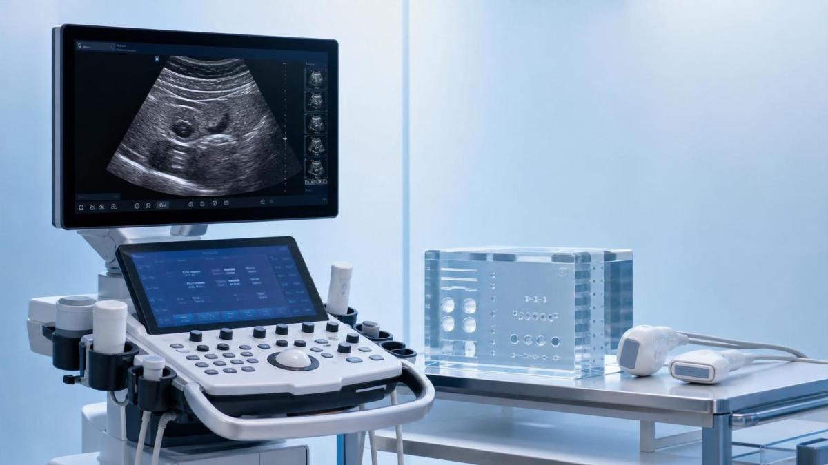
For cross-vendor comparison, accurate ultrasound image assessment should test consistency, not just peak performance. A single impressive screenshot rarely tells the full story.
The metrics that define reliable image quality
Several core metrics anchor accurate ultrasound image assessment. They do not replace clinical interpretation, but they establish whether a system is technically trustworthy.
Spatial resolution and detail separation
Axial resolution reflects how well the system separates structures along the beam path. Lateral resolution shows separation across the beam. Elevational performance affects slice thickness and boundary clarity.
Weakness in any of these areas can blur lesion margins, vessel walls, or tissue interfaces. That directly affects confidence in comparative review.
Contrast resolution and grayscale discrimination
Not all clinically relevant targets are sharply outlined. Many depend on subtle echogenic differences. Good contrast resolution helps preserve low-contrast visibility without creating false texture enhancement.
This metric becomes especially important when evaluating abdominal, vascular, and soft tissue imaging where faint differences carry diagnostic weight.
Signal-to-noise ratio and uniformity
Noise can imitate anatomy, obscure weak echoes, or inflate apparent sharpness after processing. Signal-to-noise ratio should be reviewed alongside field uniformity across depth and width.
If one part of the image is systematically cleaner than another, the issue may involve beam profile, gain compensation, or transducer element performance.
Penetration and depth-dependent stability
Penetration is not simply the deepest visible echo. The better question is whether useful contrast and recognizable structure remain at depth under realistic settings.
Accurate ultrasound image assessment should examine how image integrity changes as depth increases, especially when attenuation rises.
Temporal resolution
Frame rate affects how faithfully motion is rendered. Cardiac, fetal, vascular, and interventional imaging all depend on stable temporal performance.
Aggressive processing may improve still-image appearance while reducing responsiveness. Review should capture both static and dynamic behavior.
Artifacts are not minor flaws
Artifacts are often treated as visual nuisances, yet they are central to accurate ultrasound image assessment. They expose the limits of the system and the conditions under which interpretation becomes less reliable.
Some artifacts are physics-driven and unavoidable. Others are amplified by beam steering, gain imbalance, harmonics, or software post-processing. The review task is to tell them apart.
Artifacts that deserve close attention
- Reverberation, which can imitate repeated interfaces or contaminate near-field detail.
- Shadowing, which may be useful diagnostically but can also hide adjacent structures.
- Enhancement, which may exaggerate fluid boundaries or alter perceived contrast balance.
- Mirror image and refraction, which can create misleading duplicated anatomy.
- Speckle distortion, clutter, and side-lobe effects, which reduce confidence in subtle findings.
A strong system does not eliminate every artifact. It manages them predictably, and it allows the user to recognize when the image remains trustworthy.
Where review criteria often fail in real comparisons
Many evaluations rely on showroom settings, handpicked cases, or vendor-optimized presets. That approach can hide performance tradeoffs that emerge later in daily use.
A more defensible review framework looks at repeatability, preset transparency, operator dependency, and probe-specific behavior. It asks whether two operators can reach similar conclusions under controlled conditions.
This matters for procurement as much as for clinical operations. In value-based selection, image quality is tied to maintenance intervals, training burden, software update stability, and the likelihood of future revalidation.
Useful review criteria include
- Performance across probes rather than one flagship transducer.
- Behavior across shallow, medium, and deep targets.
- Consistency before and after software updates.
- Clarity of parameter traceability for audit or regulatory review.
- Phantom-based evidence paired with real-use imaging scenarios.
That mix reflects the kind of engineering truth emphasized by VSM. It connects measurable outputs to procurement confidence without turning technical review into brand narrative.
Applying accurate ultrasound image assessment across settings
The same framework supports several business and technical decisions, but the emphasis changes with context.
Across these settings, accurate ultrasound image assessment is less about finding a perfect image and more about proving dependable behavior within defined limits.
A practical way to strengthen decision quality
In practical terms, the strongest evaluations combine objective metrics, artifact analysis, and controlled review criteria in one workflow. Any one of those alone can mislead.
Start with a clear use case. Then define which probes, depths, targets, and motion conditions matter most. Capture baseline metrics under traceable settings. Review artifacts in context rather than treating them as isolated image defects.
Most importantly, compare systems under the same protocol. That is the only way accurate ultrasound image assessment can support defensible selection, internal validation, or performance claims.
When the next review cycle begins, it is worth turning screenshots into evidence: map the required scenarios, define pass-fail thresholds, and look closely at where image quality changes under pressure. That is usually where the most useful differences appear.
Recommended News
- 2026.10.15Medical Packaging Heat Seal Testing: Seal Strength vs. Seal Integrity TestsDr. Julian Rossi (RehabTech Specialist)
- 2026.10.14Clinic Supplier Comparison: Evaluating Cost, Delivery Reliability, and SupportDr. Alistair Thorne (Senior Biomedical Engineer)
- 2026.10.13Engineering Performance Testing Services: Evaluating Test Methods and AccuracyDr. Alistair Thorne (Senior Biomedical Engineer)
The VitalSync Intelligence Brief
Receive daily deep-dives into MedTech innovations and regulatory shifts.